| Home | E-Submission | Sitemap | Editorial Office |  
top_img
J Korean Soc Laryngol Phoniatr Logop > Volume 33(2); 2022 > Article
후두에서 기원한 IgG4 연관 가성 종양 1예

Abstract

IgG4-related disease is a fibroinflammatory condition by infiltration of IgG4-positive plasma cells that often presents as a tumorous lesion. This disease can affect nearly every organ system. After the pancreas, the head and neck region is second most common site for presentation of IgG4-related disease such as Mikulicz’s disease, Küttner tumor. The involvement of IgG4-related disease in laryngeal lesions is extremely rare. We have experienced a case of IgG4-related disease with pseudotumor formation in the larynx that is suggestive of malignancy in radiologic findings. But the pathology findings was finally confirmed as IgG4-related disease. Oral treatment with prednisolone was initiated, and the edematous mass reduced in size without permanent functional impairment of vocal fold mobility. We report our experience with a literature review.

서 론

IgG4 연관 질환(IgG4-related disease)은 전신에 침범 가능하며, 면역세포의 침윤에 의해 조직의 경화 및 염증 상태를 유발하여 종물과 유사한 형태를 보일 수 있다. Hamano 등[1]은 자가면역성 췌장염(autoimmune pancreatitis)과 혈청 IgG4 수치와의 관계를 연구하는 과정에서 처음으로 IgG4 연관 질환에 대해 보고하였으며, 췌장 외에도 경화성 담관염(sclerosing cholangitis), 후복막섬유증식증(retroperitoneal fibrosis), 폐의 염증성 위종양(inflammatory pseudotumor) 등의 형태로도 나타날 수 있다. 침범 부위는 췌장 다음으로 두경부 영역에서 흔하며 악하선, 눈물샘 및 이하선에서 주로 나타나는 것으로 알려져 있다[2]. 그 외 후두에도 발생 가능하나 그 빈도는 매우 드물며[3], 후두에서 기원한 IgG4 연관 질환은 아직까지 국내에서 보고된 바가 없다.
최근 저자들은 우측 이상와(pyriform sinus)에서 기원하여 양측 피열후두개주름(aryepiglottic fold)까지 침범하는 종양의 형태를 보이는 환자를 경험하였고, 이에 시행한 현수 후두경하 조직 검사상 IgG4 연관 가성 종양으로 최종 진단된 1예를 경험하였기에 문헌 고찰과 함께 보고하는 바이다.

증 례

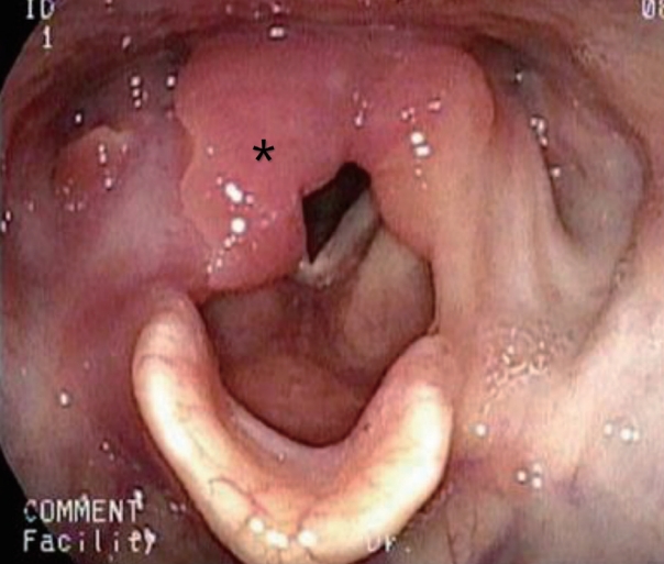
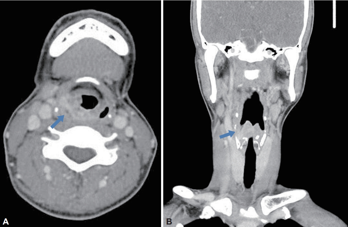
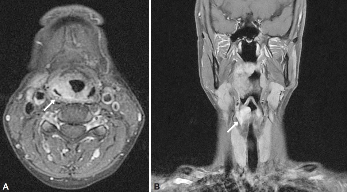
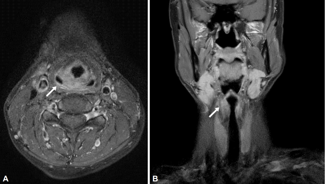
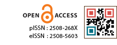
46세 여자 환자가 내원 2주 전 시행한 위내시경에서 발견된 후두의 종물을 주소로 내원하였다. 환자는 내원 한달 전부터 목 이물감이 있었고 이외 호흡곤란, 인후통, 목소리 변화 등은 없었다. 신체진찰상 비강, 경부에서는 특이 소견을 보이지 않았으나 후두내시경 상에서 우측 이상와 내측부에서 양측 피열후두개주름에 걸쳐 미만성 후두 종대가 관찰되었고 성대의 움직임은 정상 소견을 보였다(Fig. 1). 진단을 위해 시행한 경부 전산화단층촬영에서 우측 성문부에서 시작하여 일부 하인두 부근까지 침범하는 경계가 불명확한 약 2.3×1.0×1.5 cm 크기의 균일하게 조영 증강되는 종괴가 관찰되었다(Fig. 2). 경부 자기공명영상에서도 같은 위치에서 T1 강조영상에서 고신호 강도를 보이는 종괴가 관찰됐으며, 우측 피열후두개주름에서 하인두후벽(posterior hypopharyngeal wall)을 통해 좌측 피열후두개주름까지 연장하는 소견이 보였다(Fig. 3). 이외 경부에서 병적 림프절 비대는 관찰되지 않았고 악하선, 이하선에서 조영 증강되는 종괴의 소견은 없었다. 이에 성문부를 침범한 하인두암, 자가면역질환의 후두 침범, 아밀로이드증(amyloidosis) 등의 감별을 위해 현수 후두경하 조직검사를 실시하였으며 우측 이상와, 우측피열연골(arytenoids cartilage) 부위의 여러 곳에서 동결절편 검사를 실시하였으나 다수의 염증세포만 관찰되었고 악성세포는 보이지 않았다. 이후 병변으로부터 CO2 레이저를 이용하여 충분한 절제 연을 확보하여 종괴 절제를 시행하였고 지혈 후 수술을 마무리 하였다.
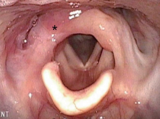
술 후 병리조직검사상 광범위한 림프형질세포 침윤(lymphoplasmacystic infiltration)이 관찰됐으며(Fig. 4A), IgG4 염색에서 양성소견을 보였다(Fig. 4B). 면역조직화학검사상 IgG 및 IgG4에서 양성을 보이는 형질세포(plasma cell)가 고배율 시야에서 300개 이상 관찰되었으며 IgG4/IgG의 비율이 0.5 이상 소견을 보여 IgG4 연관 가성종양으로 진단되었다. 추가로 시행한 복부 및 흉부 전산화단층촬영에서 다른 장기로의 침범 소견은 관찰되지 않았다. 술 후 1주일째 시행한 후두 내시경상 성대 움직임에 이상없이 수술부위는 잘 회복되고 있었고, 술 후 한달 째 시행한 후두내시경 상에서도 재발의 증거 없이 잘 회복된 소견이 관찰되었다(Fig. 5). IgG4 연관 가성종양에 대하여 3주간의 고용량 스테로이드 치료(Prednisolone 40 mg QD) 후 스테로이드 용량을 점진적으로 감량하여 지속 복용 중이며, 술 후 6개월 뒤 시행한 경부자기공명영상에서 스테로이드 복용 전 관찰됐던 고신호 강도를 보이는 종괴는 호전되었음을 확인하였고, 1년 이상 재발의 증거 없이 본원에서 추적 관찰 중이다(Fig. 6).

고 찰

Hamano 등[1]이 자가면역 췌장염 환자의 혈청 내 고농도의 IgG4 및 췌장의 면역조직화학검사상 IgG4 양성의 형질세포 침윤이 관찰된다고 처음으로 보고하였다. 그 후로 여러 연구들에서 췌장 외에도 후복막강, 폐, 눈물샘, 침샘, 대동맥 등에서도 광범위한 IgG4 양성의 형질세포들의 침윤 및 염증성 가성종양을 특징으로 하는 사례들이 보고되면서 침범된 기관에 관계없이 조직의 림프형질세포 침윤 및 경화, 환자의 혈청 IgG4 증가 등의 특징들을 보이는 질환들을 IgG4 연관 질환의 하나로 생각하는 증후군의 개념으로 자리잡았다. IgG4 연관 질환은 전신에 침범 가능하나 췌장에서 가장 많이 발생하며 다음으로 두경부 영역이 흔하며, IgG4 연관 질환 중 하나인 Ku¨ttner 종양은 주로 악하선에서, Mikulicz 병은 눈물샘 및 이하선에서 발생하게 된다. 그 외 인후두에도 발생 가능하나 그 빈도는 매우 드물며, 현재까지 보고된 사례가 소수에 불과하다[2,4-6].
Umehara 등[7]에 의해 제시된 IgG4 연관 질환에 대한 진단 기준을 살펴 보면 1) 하나 이상의 장기에 광범위한 또는 국소적인 종창 및 종물이 임상적으로 관찰되며, 2) 혈청 내 IgG4 농도가 증가해 있으며(≥135 mg/dL), 3) 뚜렷한 림프구와 형질세포의 침윤과 섬유화가 관찰되고 IgG4+와 IgG+의 비가 40% 이상이거나 IgG4+ plasma cells/HPF가 10 이상이어야 한다. 세 가지 조건을 모두 충족시킬 경우 확진(definite)할 수 있으며, 1)과 3) 조건을 만족시킬 때 추정(probable), 1)과 2) 조건을 만족시킬 때 의심(possible) 할 수 있다고 제시했다. 본 증례에서는 첫 번째와 세 번째 조건을 만족하여 IgG4 연관 질환을 추정진단 할 수 있다.
IgG4 연관 질환은 스테로이드가 1차 치료이나 재발하는 경우, 장기간의 스테로이드 복용이 필요한 경우에 리툭시맙(rituximab), 아자티오프린(azathioprine), 메토트렉세이트(methotrexate) 등의 면역억제제도 쓸 수 있다[8,9]. 하지만 IgG4 연관 질환의 침범 범위, 재발 여부, 유지 용법과 관련하여 정립된 치료 가이드라인은 아직까지 부족한 상황으로, 치료제의 선택 및 스테로이드 복용법에 대하여 주치의의 임상적 판단에 따라 치료제를 선택하며 용량 및 복용 기간을 정하게 된다[10]. 또한 IgG4 연관 질환은 재발이 흔하며 다른 부위로 침범할 수 있어 치료 중단 후에도 주기적인 영상검사와 함께 장기적 추적 관찰이 필요한 질환이다.
본 증례의 경우 성문상부에 침범한 경우로 후두내시경상에서 성대 움직임의 제한이나 기도 폐색 소견은 관찰되지 않았지만 Matsushima 등[4]은 성문부 및 성분하부까지 침범하여 기도 폐색을 유발한 IgG4 연관 후두 가성 종양 사례에 대해 보고한 바 있으며, Shaib 등[6]은 후두에서 기원한 IgG4 연관 질환으로 발생한 영구적인 성대의 마비에 대해 보고한 바 있다. 일반적으로 IgG4 연관 질환의 예후는 양호한 것으로 보고되고 있으나 Shaib 등[6]은 발병 초기 스테로이드와 같은 적절한 치료가 이루어지지 않을 경우 만성 염증과 섬유화의 진행으로 인해 성대 마비와 같은 기관의 장애가 발생하게 되며, 이는 추가적인 스테로이드 치료에도 불구하고 비가역적 손상을 유발 할 수 있다고 보고하였다[6,11]. 따라서 후두에서 발생한 IgG4 연관 질환은 진단과 함께 조기 치료가 중요하다. 하지만 보고된 사례가 소수에 불과하고 Behzadi 등[2]이 보고한 두경부 영역의 IgG4 연관 질환에 대한 영상적 특징에 대한 체계적 고찰에 따르면, 후두에서 발생한 경우 경부 전산화단층촬영상 균일한 조영 증강을 보이는 점막하 종괴를 특징으로 하지만, 증례의 수가 충분치 않아 진단적 검사로는 제한적으로 후두의 종물을 주소로 내원한 환자에서 IgG4 연관 질환을 의심하고 진단하기는 쉽지 않은 일이다[2,3]. 본 증례에서도 경부 전산화단층촬영 및 자기공명영상 상에서 하인두암을 시사하는 소견을 보였으나 후두내시경 및 임상적으로는 아밀로이드증 또는 전신성 홍반성 낭창(systemic lupus erythematous)과 같은 자가면역질환의 후두 침범 등의 감별이 필요할 것으로 사료되어 진단을 위해 조직검사를 진행하였으며, 면역조직화학검사 결과에서 IgG4 양성인 형질세포의 조직 침착 소견이 보여 IgG4 연관 후두 가성 종양으로 진단할 수 있었다.
본 증례는 후두의 종양을 주소로 내원한 환자에서 시행한 전산화단층촬영 및 자기공명영상 등과 같은 영상의학적 검사뿐만 아니라 임상적 판단 및 후두내시경 소견 역시 중요함을 시사하고 있으며, 발병률은 매우 드문 것으로 알려져 있지만 IgG4 연관 질환 역시 가성 종양의 형태로 후두에서 발생 할 수 있음을 숙지하여 조직검사 및 조기 치료를 통해 IgG4 연관 질환으로 인한 만성 염증 및 섬유화로 발생할 수 있는 비가역적인 성대의 손상을 예방하는 것이 중요하겠다. 이에 저자들은 조기에 후두에서 발생한 가성 종양을 진단하고 성공적으로 치료하였기에 문헌고찰과 함께 보고하는 바이다.

ACKNOWLEDGEMENTS

None.

NOTES

Conflicts of Interest

The authors have no financial conflicts of interest.

Authors’ Contribution

Conceptualization: all authors. Data curation: all authors. Formal analysis: all authors. Funding acquisition: all authors. Investigation: all authors. Methodology: all authors. Project administration: all authors. Resources: all authors. Software: all authors. Supervision: all authors. Validation: all authors. Visualization: all authors. Writing—original draft: all authors. Writing—review & editing: all authors. Approval of final manuscript: all authors.

Fig. 1.
Laryngoscope findings. Diffuse laryngeal swelling mimicking hypopharyngeal cancer formed primarily in the right arytenoid region (astrix).
jkslp-2022-33-2-110f1.jpg
Fig. 2.
Preoperative axial (A) and coronal (B) CT image. A mass like lesion measuring 2.3×1.0×1.5 cm with contrast enhancing in the periphery of the right arytenoid region (blue arrow).
jkslp-2022-33-2-110f2.jpg
Fig. 3.
Preoperative axial (A) and coronal (B) MRI image. T1-weighted image shows high signal intensity mass occupying from right pyriform sinus to left aryepiglottic fold through posterior hypopharyngeal wall (white arrow).
jkslp-2022-33-2-110f3.jpg
Fig. 4.
Pathologic findings. Immunohistochemical staining shows diffuse lymphoplasmacystic infiltration (H&E, ×400) (A). Immunoglobulin G4 (IgG4)-positive plasma cells, measuring up to 20/HPF. The black arrow indicates a plasma cell (IgG4, ×400) (B).
jkslp-2022-33-2-110f4.jpg
Fig. 5.
Postoperative laryngoscope findings. The pseudotumor was reduced in size after administration of prednisolone compared to preoperative laryngoscope findings (astrix).
jkslp-2022-33-2-110f5.jpg
Fig. 6.
Post-treatment axial (A) and coronal (B) MRI. The high signal intensity mass seen in the preoperative MRI was improved after administration of prednisolone (white arrow).
jkslp-2022-33-2-110f6.jpg

REFERENCES

1. Hamano H, Kawa S, Horiuchi A, Unno H, Furuya N, Akamatsu T, et al. High serum IgG4 concentrations in patients with sclerosing pancreatitis. N Engl J Med 2001;344(10):732-8.
crossref pmid
2. Behzadi F, Suh CH, Jo VY, Shanmugam V, Morgan EA, Guenette JP. Imaging of IgG4-related disease in the head and neck: A systematic review, case series, and pathophysiology update. J Neuroradiol 2021;48(5):369-78.
crossref pmid
3. Yoon HJ, Park WS, Ji YB, Lee SH. Two cases of immunoglobulin G4-related sclerosing disease mimicking nasopharyngeal carcinoma. Korean J Otorhinolaryngol-Head Neck Surg 2016;59(4):304-8.
crossref
4. Matsushima K, Ohira S, Matsui H, Fukuo A, Honma N, Wada K, et al. IgG4-related disease with pseudotumor formation in the larynx. Auris Nasus Larynx 2020;47(2):305-8.
crossref pmid
5. Mulholland GB, Jeffery CC, Satija P, Côté DW. Immunoglobulin G4-related diseases in the head and neck: A systematic review. J Otolaryngol Head Neck Surg 2015;44(1):24.
crossref pmid pmc pdf
6. Shaib Y, Ton E, Goldschmeding R, Tekstra J. IgG4-related disease with atypical laryngeal presentation and Behçet/granulomatous polyangiitis mimicking features. BMJ Case Rep 2013;2013:bcr2013009158. Shaib Y, Ton E, Goldschmeding R, Tekstra J. IgG4-related disease with atypical laryngeal presentation and Behçet/granulomatous polyangiitis mimicking features. BMJ Case Rep 2013;2013:bcr2013009158.
pmid pmc
7. Umehara H, Okazaki K, Masaki Y, Kawano M, Yamamoto M, Saeki T, et al. Comprehensive diagnostic criteria for IgG4-related disease (IgG4-RD), 2011. Mod Rheumatol 2012;22(1):21-30.
crossref pmid
8. Khosroshahi A, Wallace ZS, Crowe JL, Akamizu T, Azumi A, Carruthers MN, et al. International consensus guidance statement on the management and treatment of IgG4-related disease. Arthritis Rheumatol 2015;67(7):1688-99.
crossref pmid pdf
9. Lee CM, Alalwani M, Prayson RA, Gota CE. Retrospective single-centre analysis of IgG4-related disease patient population and treatment outcomes between 2007 and 2017. Rheumatol Adv Pract 2019;3(1):rkz014.
crossref pmid pmc
10. Khosroshahi A, Wallace ZS, Crowe JL, Akamizu T, Azumi A, Carruthers MN, et al. International consensus guidance statement on the management and treatment of IgG4-related disease. Arthritis Rheumatol 2015;67(7):1688-99.
crossref pmid pdf
11. Shimizu Y, Yamamoto M, Naishiro Y, Sudoh G, Ishigami K, Yajima H, et al. Necessity of early intervention for IgG4-related disease--delayed treatment induces fibrosis progression. Rheumatology (Oxford) 2013;52:679-83.
crossref pmid
Editorial Office
Journal of The Korean Society of Laryngology, Phoniatrics and Logopedics, Ewha Womans University, College of Medicine.
Anyangcheon-ro 1071, Yangcheon-gu, Seoul, Republic of Korea
TEL: +82-10-3008-3075   FAX: +82-2-2646-3076    E-mail: secretary@kslpl.org
About |  Browse Articles |  Current Issue |  For Authors and Reviewers
Copyright © Journal of The Korean Society of Laryngology, Phoniatrics and Logopedics.                 Developed in M2PI