성대진동검사의 최신 지견
Recent Advances in Examination of Vocal Fold Vibration
Article information

Trans Abstract
Human vocal cords vibrate as quickly as 100–250 times per second, so it is impossible to observe them with normal endoscopic diagnostic equipment. High-speed videolaryngoscopy (HSV) allows the visualization of non-periodic vibratory motion of vocal fold beyond the limitation of videostroboscopy. New developed post-processing methods that converts HSV to two-dimensional videokymography (2D VKG) using U-medical image-processing software can provide quantitative information on vocal fold mucosa vibration. Multifunctional laryngeal examination system is composed of 3 kinds of examinations such as HSV, 2D scanning digital kymography (2D DKG) and line scanning digital kymography (DKG). Evaluation of entire vocal cord vibratory pattern in each cord is possible using 2D DKG and a faster and more reliable quantitative information can be obtained. As this system is used in clinical and research, it is expected to bring much advances to the diagnosis of voice disorders. In this review, I will introduce the principles and advantages on examination of the vocal fold vibration, which is in the spotlight recently, and proceed with the literature review.
서론
사람의 성대는 1초에 100~250회 정도로 빠르게 진동하기 때문에 정상적인 내시경 진단장비로는 관찰하기가 불가능하다. 1895년 Oretel이 탈봇의 원리를 이용하여 성대 진동을 느린 화면으로 관찰한 이래로 많은 검사 기구들이 개발되어 성대의 진동을 관찰하려는 시도가 현재에도 진행되고 있다[1-3]. 현재까지도 임상에서 가장 많이 사용되는 후두 스트로보스코피(laryngeal stroboscopy)가 있지만 심한 애성이 있어 3~5초 이상의 안정적인 발성이 어려운 경우는 검사가 부적합하다.
이때 사용할 수 있는 방법이 초당 2000~5000 프레임으로 성대의 진동을 관찰할 수 있는 초고속 비디오 후두내시경 검사(high-speed videolaryngoscopy, HSV)이다. 뿐만 아니라 초고속 영상을 촬영한 뒤 이미지 처리과정을 통하여 얻을 수 있는 카이모그래피(kymography)는 성대의 불규칙한 진동의 관찰도 가능하게 한다[1]. 카이모그래피는 비디오 카이모그래피(videokymography, VKG)와 디지털 카이모그래피(digital kymography, DKG)로 나눌 수있다. 비디오 카이모그래피는 비디오 카메라만으로 측정한 영상으로, 라인 스캔 비디오 카이모그래피와 평면 스캔 비디오 카이모그래피가 있으며, 디지털 카이모그래피는 초고속 비디오 내시경 영상을 촬영한 뒤, 이미지 처리 과정을 통하여 비디오 카이모그래피와 거의 유사한 영상을 얻는 방식이며, 이 방식에도 라인 스캔 디지털 카이모그래피와 평면 스캔 디지털 카이모그래피가 있다. 이러한 초고속 후두내시경 영상을 이용한 평면 스캔 디지털카이모그래프(2D DKG) 영상 생성을 통하여 위상대칭지수(Phase Symmetry Index), 진폭대칭지수(Amplitude Symmetry Index), 개방지수(open quotient), 폐쇄지수(close quotient) 등의 성대 진동의 정량적인 정보를 얻을 수 있었다[2]. 또한, 최근에 새롭게 개발된 초고속 디지털 카메라로 촬영한 영상을 이용하여 DKG와 2D DKG를 동시에 획득함으로써 다양한 형태의 이미지를 실시간으로 동시에 제공할 수 있었고, 형태학적 및 기능적 진동 패턴도 분석할 수 있었다[3].
최근에는 앞에서 언급한 고속 비디오 후두 내시경 검사, 2D 디지털 카이모그래피 및 라인 스캐닝 디지털 카이모그래피를 실시간으로 동시에 검사가 가능한 혁신적인 기술인 다기능 후두성능검사시스템(multifunctional laryngeal examination system)이 개발되어 종합적으로 성대 진동을 관찰할 수 있게 되었으며, 이는 다양한 성대 진동 장애 병변의 진단에 이용될 예정이다.
본 논문에서는 여러 가지 최신의 성대 진동 검사들에 대해 알아보고자 한다.
본론
최근에 많이 이용되고 있는 여러가지 성대진동검사에 대해 알아보고자 한다.
후두 스트로보스코피(Laryngeal stroboscopy)
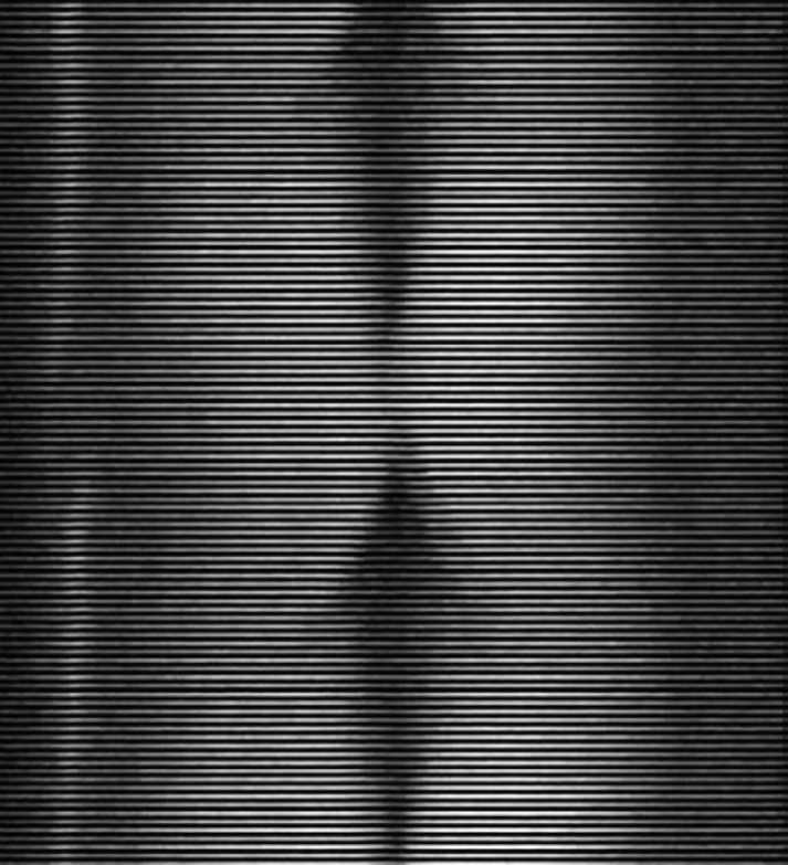
현재 사용되고 있는 후두 스트로보스코피는 전기 전자 기술의 발달로 성대를 직접 눈으로 관찰하는 것이 아니라 내시경과 Charge Coupled Device(CCD) 카메라를 통해 들어온 데이터가 모니터로 출력되는 영상에서 성대의 느린 움직임을 관찰한다. 2010년도에 발표된 연구에 의하면 실제의 움직임은 없으나 움직임이 있는 것으로 인식되는(최소한 17 Hz 이상으로 디스플레이가 가능한) 가현 운동(apparent motion) 지각과 화면에 디스플레이 되는 영상을 주사할 때 깜빡임 현상이 나타나는 플리커(flicker) 현상이 서로 다른 시지각(visual perception) 현상으로 이러한 원리가 최근 스트로보스코피의 원리로 인정받고 있다[4]. 최소 50 Hz 이상의 속도로 영상을 주사해야만 화면의 깜박임을 눈으로 지각하지 못한다는(flikerless) 원리가 내포되어 있다고 할 수 있다. 오랫동안 후두 스트로보스코피를 구현하는 방식으로 기본주파수에 맞게 단속 광원을 조절하여 스트로보 효과를 이용하는 방법을 이용하여 왔다[5]. 이는 대부분의 스트로보스코피가 이용하는 방법인데, 진동하는 성대를 촬영할 때 성대 진동 주기와 동기화된 고속 발광 단속 광원을 비춰주면 나타나는 영상은 성대의 실제 움직임이 아니라 발광되는 순간의 성대의 모습이 느리게 관찰된다. 이때 후두 스트로보스코피의 동기화 신호 추출은 전기성문파(electroglottography) 신호를 이용하는 방식이 가장 널리 사용되고, 그 외 음성 혹은 진동검출기를[6] 이용하는 방식이 소개되기도 하였다. 다른 방법으로는 기본 주파수를 이용하여 셔터스피드를 조절하여 단속 광원이 아닌 연속 광원을 이용하여 스트로보 효과를 나타내는 방법이다. 2000년도에 들어 여러 연구에서 연속 광원을 이용하여 셔터 스피드를 성대 진동과 동기화하면 새롭게 스트로보 효과를 구현할 수 있다고 보고하였다[7]. 하지만, 특정 후두에 대한 시각적이고 객관적인 평가는 스트로보스코피 이미징을 사용하여서는 신뢰할 수 없어서 후두 고속 비디오 내시경 검사 및 기타 대체 이미징 방식을 일상적인 임상 실습에 통합하여 음성 장애 관리를 향상시키는 추가 연구가 필요하다[7]. Fig. 1은 초당 30 프레임(fps)을 캡처하여 각 진동주기의 다른 지점에서 이미지를 모아서 편집한 성대 진동의 슬로우 모션을 보여주는 스트로보스코피 영상이다.
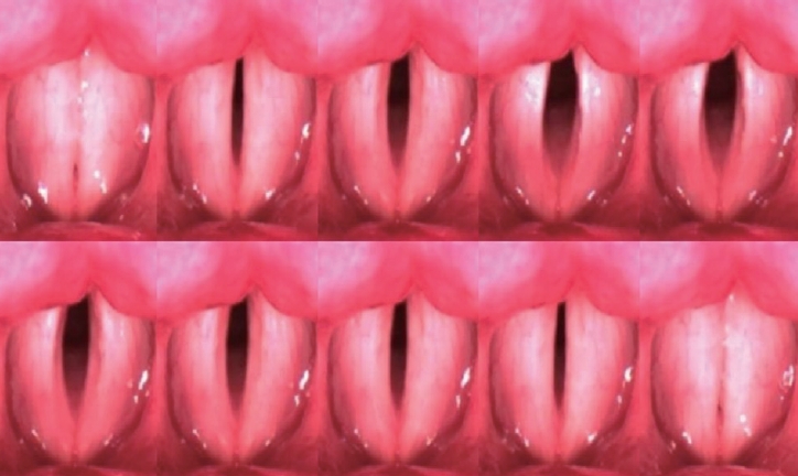
Montage of stroboscopic images obtained from successive points in several glottal cycles.
초고속 비디오 후두내시경 검사(High-speed videolaryngoscopy)
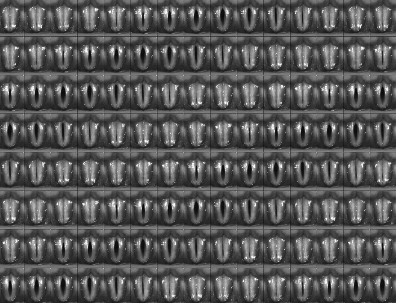
1990년대 연구목적으로 도입된 초고속 비디오 후두내시경 검사(HSV)는 성대 진동 운동의 관찰을 위한 가장 정확한 도구로 간주되었다. 그러나 상업적 HSV 시스템은 해결되지 않은 기술적, 평가방법론적 한계와 HSV의 유효성 및 임상적 관련성에 관한 정보 부족으로 인해 임상적으로는 널리 사용되지 않았다[8]. HSV로 촬영하여 얻는 초고속 디지털 영상(high-speed digital imaging, HSDI)은 최근에 가장 널리 사용되는 점막 운동의 시각화 방법이 되었다. 후두 스트로보스코피 검사로 생성된 착시와는 달리 HSDI의 높은 프레임 속도는 점막파를 상세하게 시각화 하여 단일 성대 주기에서 여러 이미지를 캡처한다[9]. Fig. 2는 정상 성인 남성에서 발성 시 초당 3000 프레임으로 촬영된 성대의 HSV 영상소견이다[2]. 최근의 보고에 의하면 HSV 데이터를 분석하는 데 사용된 방법이 다양한 성대 병리를 가진 환자의 성대 진동의 특성을 문서화하고 진동장애의 심각성을 추정하기 위해 진동 장애를 조사하는 데 매우 유용할 수 있음을 입증했다[10-13]. 대부분의 초고속 비디오 후두내시경 검사는 라인 스캐닝 카이모그래피 기능을 사용할 수 있도록 소프트웨어를 제공해 주므로 여러 주기에 걸쳐 점막파동 및 주기성, 대칭성을 시각적으로 빠르게 관찰할 수 있어 HSV와 병행하여 효율적으로 사용되고 있다. 2017년 보고에서 성대 진동의 모든 단계에 대한 직렬 HSV 이미지를 U-medical 이미지 처리 소프트웨어를 사용하여 스캐닝 방법에 의해 변환시켰는데. 이러한 후 처리된 2D DKG 이미지는 다양한 진동 위상을 포함하여 성대 점막 진동에 대한 정량적 정보를 제공할 수 있었다[2].
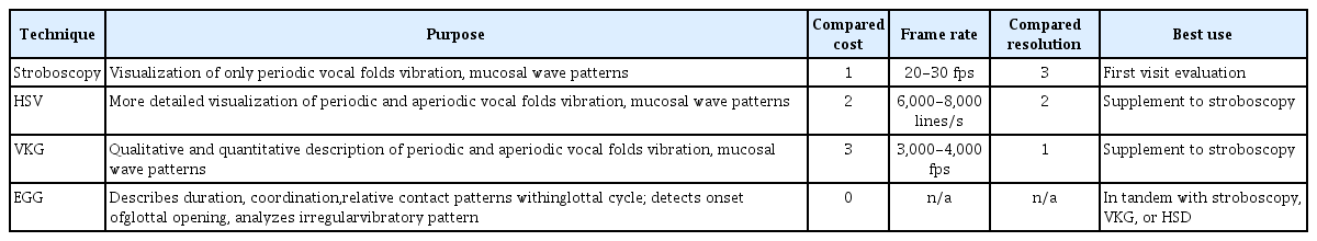
새롭게 획득된 2D DKG와 DKG 사이의 진폭 대칭 지수, 위상 대칭 지수, 개방 지수 및 폐쇄 지수의 차이를 분석하였고 2D DKG와 DKG에서 성대 진동 운동의 정량적 파라미터 사이에는 통계적 차이가 없다는 결과를 보여주었다[2]. HSDI에 의해 기록된 더 높은 프레임 속도와 많은 이미지 수는 분석에 더 많은 시간과 비용이 소요되지만 HSDI에 의해 생성된 시각화는 주파수에 독립적이므로 비주기적인 병리학적 성대의 진동을 진단하는 데는 더 효과적이다[9]. Table 1은 여러가지 성대 진동 검사 기기의 차이점을 보여주고 있다. Sekimoto 등[14]은 1200 fps의 기록 속도로 비교적 저렴한(3000 달러) 고속 촬영 시스템이 제안되었지만 상대적으로 해상도가 낮은 단점이 지적되었다.
Comparison of available current mucosal wave imaging techniques
카이모그래피(Kymography)
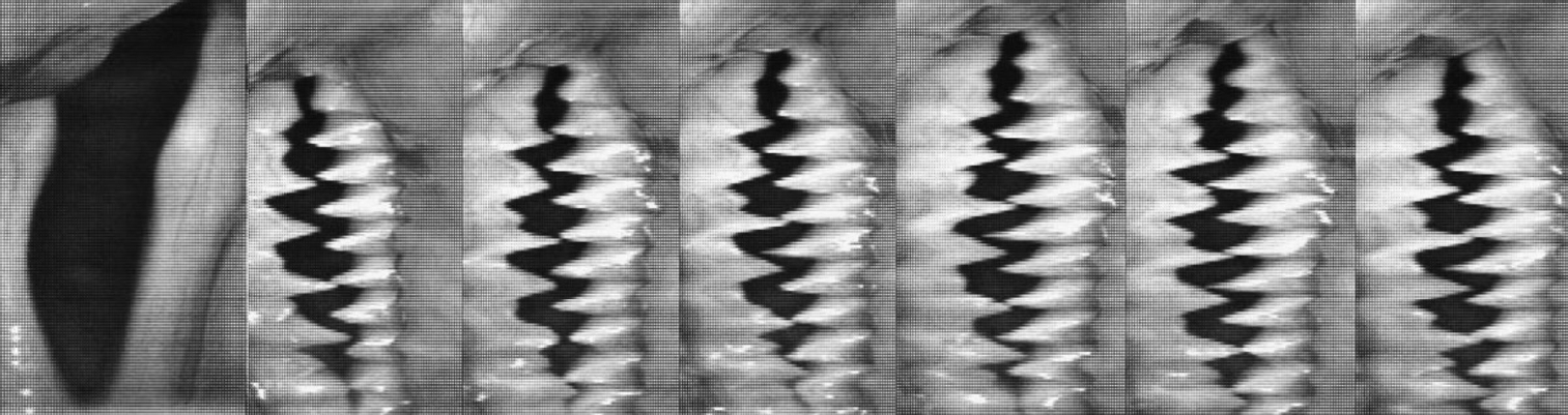
디지털 카이모그래피(DKG)의 출현과 함께 HSDI는 시스템이 대폭 개선되었다. 이는 성대 진동을 보다 효과적으로 연구하는 능력과 카이모그램을 통한 점막 진동 파라미터와 진동을 정량화하는 능력이 도입되었기 때문이다. DKG는 2000~5000 fps 속도로 HSDI가 얻은 풀 프레임 이미지를 사용하고 컴퓨터 소프트웨어로 분석한다[8,9,15,16]. 이 소프트웨어는 비디오의 연속 프레임 각각에서 추출된 성문 축(glottal axis)에 직각인 픽셀 라인을 사용자가 원하는 대로 선택할 수 있다(Fig. 3A). 영상 저장 후 나오는 라인 추출은 픽셀 라인이 선택되기 전에 각도 보정 및 후두개 또는 피열 연골에 의한 카메라 시야의 막힘 여부 확인을 가능하게 해주고 픽셀 라인이 한번 추출되면, 프레임 번호에 기초하여 연속적으로 나란히 구성된다. 이것은 점막 파의 움직임을 시각화 하여 개방 및 폐쇄 위상, 주기성, 좌우 대칭, 위상차 및 진폭을 보여주는 카이모그램(kymogram)을 만든다(Fig. 3B)[17]. HSDI와 함께 사용되는 DKG는 HSDI만으로 프레임 단위의 성대 사이클을 분석하는 것보다 점막 파 매개 변수를 설명하는 정량적 및 정성적 결과를 얻을 수 있는 보다 빠르고 안정적인 방법을 제공한다. VKG는 HSDI와 결합된 DKG 검사 혹은 HSDI 단독 검사 보다 성대 점막 진동을 측정하는데 보다 효율적인 측정 방법이다. VKG는 DKG와 달리 녹화 전에 단일 픽셀 라인을 선택해야 하고 VKG로 기록된 단일 픽셀 라인은 HSDI 또는 DKG로 HSDI로 기록한 일반적인 풀 프레임 이미지보다 해상도가 훨씬 낮다[17]. VKG는 HSDI 만큼 많은 데이터를 기록하지 않기 때문에 CCD에 의해 기록된 하나의 픽셀 라인의 낮은 해상도의 프레임 비율은 실질적으로 증가할 수 있다. VKG에 사용되는 CCD의 프레임 속도는 거의 8000 fps에 달한다[18,19]. VKG는 HSDI 보다 높은 프레임 속도를 갖기 때문에 성대 주기 사이클 당 더 많은 사진을 기록할 수 있어 보다 정확한 카이모그램 및 점막파 매개 변수 분석이 가능하다(Fig. 4). 2016년엔 Wang 등[20]에 의해 새로운 VKG 시스템이 개발되어 전체 성대의 점막 진동 패턴과 개방 및 폐쇄 지속 시간의 검사가 용이하게 되었다. 또한 이 시스템은 진폭 및 위상의 대칭과 내측 피크 및 측면 피크의 모양을 평가할 수 있는 기능을 가지고 있어(Fig. 5) 급성 후두염 환자에서 성대의 진동을 평가하는 향상된 능력을 보여주었다(Figs. 6, 7)[20]. 최근에는 Kang 등[3]에 의해 초고속 비디오 후두내시경검사를 기반으로 하여 후두경 소견과 디지털 카이모그래피, 2D 평면 카이모그래피를 동시에 관찰할 수 있는 시스템을 개발되었다. 이 기기를 이용하면 성대 전체의 진동을 분석할 수 있었고, 특징적으로 이 시스템의 2D VKG는 초당 32400라인과 25프레임을 보여주어 한 라인만 특별하게 관찰되는 라인 스캐닝 비디오 카이모그래피와는 차별화하였다. 픽셀 수에 따라 이미지의 프레임수가 다르게 나타났고 2D DKG에서는 2픽셀 라인 스캐닝에서 정량적 평가가 유용하였다(Fig. 8)[3]. 또한 이러한 시스템을 통해 후두내시경 검사로는 정확한 관찰이 어려운 좌측 성대 반흔 환자에서 DKG와 2D VKG를 이용한 검사로 양측 성대 진동의 차이를 정확히 확인할 수 있었다(Fig. 9). 2019년도엔 Bae 등[21]이 2D DKG가 이중음성의 진단에 있어 빠른 해석이 가능하고 진단 정확도가 비교적 높았다는 보고를 하였다. 성대 폴립, 성대마비, 근긴장성 발성장애의 경우를 2D DKG로 분석한 결과 공간 점유 병변이 있는 환자의 경우 보다는 일측성 성대마비와 근긴장성 발성 장애 환자에서 의미 있는 결과를 보였다[21]. Fig. 10은 이중 음성을 가진 우측 일측성 성대마비 환자의 2D DKG 영상을 보여준다.
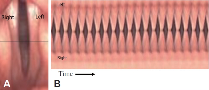
It shows the vibratory pattern of nearly periodic type. A: Image of line-scan placement at the widest section of the glottis in DKG. B: Kymogram of periodic vibration obtained using DKG. DKG: digital kymography.
VKG image of vocal folds with asymmetrical vibration. VKG: videokymography.

Characteristic VKG images generated by the new VKG system during phonation. A: The mucosal wave pattern of the entire vocal folds was captured during a single examination. B: The symmetry of the amplitude (arrow) could be assessed. C: The closing (a) and opening (b) durations could be differentiated. D, E: The shape of the medial (arrow) and lateral peaks (arrow) could be assessed. VKG: videokymography
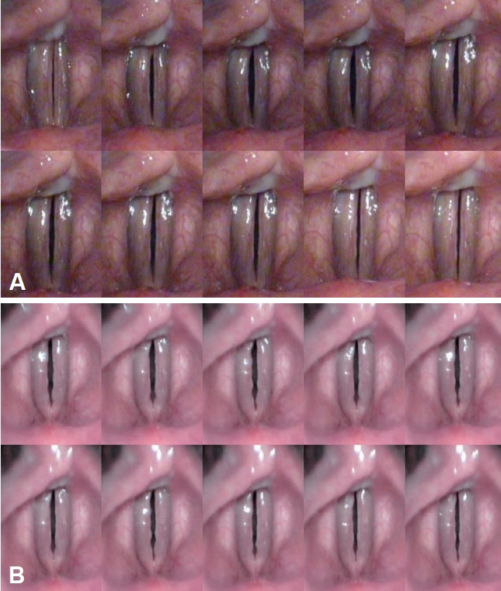
Images obtained by laryngeal videostroboscopy (A) and the new VKG system (B) at the pretreatment status. VKG: videokymography.

Imaging findings of laryngeal videostroboscopy and the new VKG system after 4 weeks. The mucosal waves of both cords show almost normal, and the shapes of their medial peaks show normal, as determined using the new VKG system. VKG: videokymography.

Examples of real-time dual DKG and 2D DKG (normal subject, 34-year-old male). A1: navigation videoendoscopy. Example of real-time 2D DKG (A2, A3: 1 pixel line, A4: 2 pixel lines, A5: 3 pixel lines, A6: 4 pixel lines, A7: 5 pixel lines). Example of real-time DKG (B1, B2: 1 pixel line, B3: 2 pixel lines, B4: 3 pixel lines, B5: 4 pixel lines, B6: 5 pixel lines). 2D DKG: two-dimensional scanning digital kymography.

Examples of real-time dual DKG and 2D DKG in patient with leftt vocal cord scar (M/50-year-old). 2D DKG: two-dimensional scanning digital kymography.
Sequential image of two-dimensional scanning digital kymography in cases with diplophonia, right vocal fold palsy (left–right cords asymmetric; male/67 years old).
다기능 후두성능검사시스템(Multifunctional laryngeal examination system)
최근에는 초고속 후두 비디오내시경 검사(HSV)를 비롯하여, 라인 스캐닝 디지털 카이모그래피 검사(Line Scanning DKG), 평면 스캐닝 디지털 카이모그래피 검사(2D DKG) 등 3가지 검사를 하나의 초고속 디지털 카메라 시스템으로 촬영하여 성대 진동을 평가할 수 있는 다기능 후두성능검사시스템이 개발되었다. 3가지 검사가 동시에(simultaneous) 가능하고 단일 모니터에서 구현되기 때문에, 검사 시간의 단축, 환자의 불편감 해소뿐 아니라 3가지 검사의 종합적 평가가 가능하여 보다 정확한 진단이 가능하게 되었고, 이러한 검사는 실시간(quasi real-time)으로 가능하여, 즉각적인 성대 진동 평가를 할 수 있다(Fig. 11). 다기능 후두성능검사시스템은 글로벌 셔터 방식의 초고속 Complementary Metal Oxide Semiconductor 디지털 카메라를 이용하여 셔터스피드를 1/2000초에서 1초까지 설정 가능하고 별도의 의료내시경과 내시경용 광원장치를 통해 성대의 진동 양상을 여러 가지 방법으로 촬영하는 시스템이다. 이러한 시스템을 이용하면 기능성 음성장애, 연축성 발성장애, 성대 진전(vocal tremor), 불완전 성대마비등의 검사 및 진단에 유용할 것으로 기대된다.

Multifunctional laryngeal examination system inncludes. A: High-speed videoendoscopy. B: 2D scanning digital kymography. C: Line scanning digital kymography. Adapted from U-medical Co. catalog 2020.
결론
음성 장애의 진단에 가장 중요한 검사는 성대 진동을 확인하는 것이다. 후두 스트로보스코피가 성대 진동 장애의 진단 도구로 사용된 이후로 많은 기구들을 개발되었고 임상에 널리 이용되고 있다. 초고속 디지털 영상 및 초고속 비디오 내시경, 이러한 영상을 이용한 평면 스캔 카이모그래피와 라인 스캔 카이모그래피, 앞의 세 가지 검사를 동시에 가능한 다기능 후두성능검사시스템 등이 임상 및 연구에 이용되면서 음성장애의 진단 영역에 많은 발전을 가져올 것으로 기대하고 있다.
Acknowledgements
This work was supported by a 2-Year Research Grant of Pusan National University.
Notes
Conflicts of Interest
The authors have no financial conflicts of interest.
Authors’ Contribution
Conceptualization: Jin-Choon Lee. Data curation: all authors. Formal analysis: all authors. Funding acquisition: Jin-Choon Lee. Investigation: all authors. Methodology: all authors. Project administration: Jin-Choon Lee. Resources: all authors. Software: Inho Bae. Supervision: Inho Bae. Validation: all authors. Visualization: all authors. Writing—original draft: Jin-Choon Lee. Writing—review & editing: all authors. Approval of final manuscript: all authors.