


ņä£ ļĪĀ
ņŗĀĻ▓Įņ┤łņóģņØĆ ņŗĀĻ▓Įņ┤łņäĖĒżņŚÉņä£ ĻĖ░ņøÉĒĢśļŖö Ēö╝ļ¦ēņ£╝ļĪ£ ļæśļ¤¼ņŗĖņŚ¼ ņĀÉļ¦ē ĒĢśņŚÉ ņŻ╝ļĪ£ ļ░£ņāØĒĢśļŖö ņ¢æņä▒ ņóģņ¢æņØ┤ļŗż[1]. ņŗĀĻ▓Įņ┤łņäĖĒżĻ░Ć ņŚåļŖö ĒøäĻ░ü ļ░Å ņŗ£ņŗĀĻ▓Į ļō▒ņØä ņĀ£ņÖĖĒĢ£ ļ¬©ļōĀ ņŗĀĻ▓ĮņŚÉņä£ ļ░£ņāØĒĢĀ ņłś ņ׳Ļ│Ā ļīĆļČĆļČä ļŗ©ņØ╝ ņóģļ¼╝ņØ┤ļŗż[2]. ļæÉĻ▓ĮļČĆņŚÉņä£ļŖö ņØĖļæÉ ņŻ╝ņ£ä Ļ│ĄĻ░ä, ĒśĆ, ņØĖļæÉ, ņ×ģņłĀ ļ░Å ņŚ░ĻĄ¼Ļ░£ ļō▒ņŚÉņä£ ļ░£ņāØ ĒĢĀ ņłś ņ׳ļŗż[2,3]. ĒøäļæÉņŚÉļŖö ļ╣äĻĄÉņĀü ļō£ļ¼╝Ļ▓ī ļ░£ņāØĒĢśĻ│Ā Ēö╝ņŚ┤ĒøäļæÉĻ░£ņŻ╝ļ”ä, Ļ░Ćņä▒ļīĆ ļ░Å ĒøäļæÉĻ░£ ļō▒ņŚÉ ļéśĒāĆļéĀ ņłś ņ׳ļŗż[4]. ņĀäĒśĢņĀü ņ×äņāü ņåīĻ▓¼ņØĆ ļČĆļō£ļ¤¼ņÜ┤ Ēæ£ļ®┤ņØä Ļ░Ćņ¦ä ņøÉĒśĢ ņóģļ¼╝ņØ┤ļ®░ ņ£ĀļæÉņ¢æ ņóģļ¼╝ņØś ņ¢æņāüņØĆ ļ¦żņÜ░ ļō£ļ¼╝ļŗż[4,5].
ņØīņä▒ ļ│ĆĒÖöņÖĆ Ļ▓ĮļČĆ ņØ┤ļ¼╝Ļ░É ļō▒ņØä ņŻ╝ņåīļĪ£ ļé┤ņøÉĒĢ£ 76ņäĖ ļé©ņ×ÉņŚÉņä£ ņŗ£Ē¢ēĒĢ£ ĒøäļæÉļé┤ņŗ£Ļ▓Į Ļ▓Ćņé¼ņŚÉņä£ Ēö╝ņŚ┤Ļ░äĻĘ╣ņŚÉ ņøÉĒśĢņØś ņ£ĀļæÉņ¢æ ņóģļ¼╝ņØ┤ Ļ┤Ćņ░░ļÉśņ¢┤ ĒøäļæÉļ»ĖņäĖņłśņłĀņØä ņŗ£Ē¢ēĒĢśņśĆĻ│Ā ņŗĀĻ▓Įņ┤łņóģņ£╝ļĪ£ ņĄ£ņóģ ņ¦äļŗ©ļÉśņŚłļŗż. ņĀĆņ×ÉļōżņØĆ ļ¦żņÜ░ ļō£ļ¼Ė ņ£ĀļæÉņ¢æ ņóģļ¼╝ņØś ņ¢æņāüņØä ļ│┤ņØĖ ĒøäļæÉ ņŗĀĻ▓Įņ┤łņóģņØä Ļ▓ĮĒŚśĒĢśņśĆĻĖ░ņŚÉ ļ¼ĖĒŚī Ļ│Āņ░░Ļ│╝ ĒĢ©Ļ╗ś ļ│┤Ļ│ĀĒĢśĻ│Āņ×É ĒĢ£ļŗż.
ņ”Ø ļĪĆ
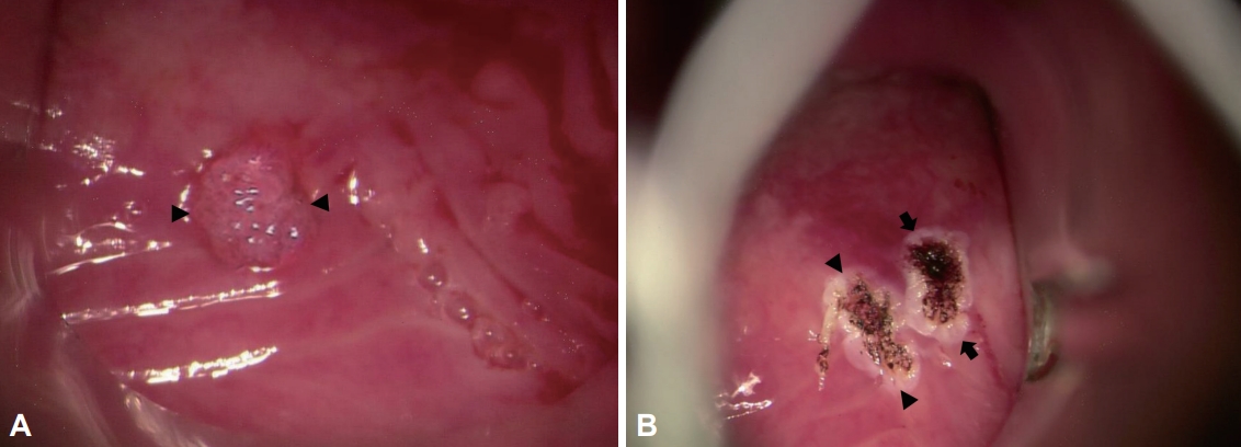
76ņäĖ ļé©ņ×É ĒÖśņ×ÉĻ░Ć ņłśĻ░£ņøö ņĀäļČĆĒä░ ņŗ£ņ×æļÉśņŚłĻ│Ā, ņĄ£ĻĘ╝ 2-3Ļ░£ņøöļÅÖņĢł ņĢģĒÖöļÉ£ ņØīņä▒ ļ│ĆĒÖöņÖĆ Ļ▓ĮļČĆ ņØ┤ļ¼╝Ļ░É ļō▒ņØä ņŻ╝ņåīļĪ£ ļé┤ņøÉĒĢśņśĆļŗż. Ļ│ĀĒśłņĢĢ, ļŗ╣ļć©, Ļ│Āņ¦ĆĒśłņ”Ø ļō▒ņØś ņ¦łĒÖśņØ┤ ņ׳Ļ│Ā ĒØĪņŚ░ļĀźņØĆ ņŚåņŚłņ£╝ļ®░ ņØīņŻ╝ļĀźņØĆ ļ»Ėļ»ĖĒĢśņśĆļŗż. Ļ░Øļŗ┤, ĻĖ░ņ╣© ļ░Å ņŚ░ĒĢśĻ│żļ×Ć ļō▒ņØĆ ĒśĖņåīĒĢśņ¦Ć ņĢŖņĢśļŗż. Ļ▓Įņä▒ ĒøäļæÉļé┤ņŗ£Ļ▓Į Ļ▓Ćņé¼ņŚÉņä£ Ēö╝ņŚ┤Ļ░äĻĘ╣ņŚÉ Ēæ£ļ®┤ņØ┤ ņÜĖĒēüļČłĒēüĒĢ£ 5├Ś3 mm Ēü¼ĻĖ░ņØś ņ£ĀļæÉņ¢æ ņóģļ¼╝ņØ┤ Ļ┤Ćņ░░ļÉśņŚłĻ│Ā, ņä▒ļīĆ ļé┤ņĀä ņŗ£ ņä▒ļ¼ĖĒŗłņØ┤ Ļ┤Ćņ░░ļÉśņŚłļŗż(Fig. 1A and B). ņä▒ļīĆņ¦äļÅÖĻ▓Ćņé¼ņŚÉņä£ ņĀÉļ¦ē ĒīīļÅÖņØĆ Ļ▓ĮĒĢśĻ▓ī Ļ░ÉņåīļÉ£ ņ¢æņāüņØ┤ņŚłĻ│Ā, ņä▒ļ¼ĖĒŗłņØ┤ Ļ┤Ćņ░░ļÉśņŚłļŗż. GRBAS ņ▓ÖļÅä ĒÅēņĀĢļ▓ĢņŚÉņä£ļŖö ļīĆļČĆļČä 1ņĀÉņØ┤ņŚłņ£╝ļ®░, ņĄ£ņן ļ░£ņä▒ ņ¦ĆņåŹņŗ£Ļ░äņØĆ 13ņ┤łļĪ£ ņĀĢņāü ļ▓öņ£äņśĆļŗż. ļŗżļ®┤ņØīņä▒Ļ▓Ćņé¼(multi-dimensional voice program, PENTAX Medical, Montvale, NJ, USA)ņŚÉņä£ ĻĖ░ļ│Ė ņŻ╝ĒīīņłśĻ░Ć 173.21 Hz, ņŻ╝Ēīīņłś ļ│ĆļÅÖļźĀ(jitter)ņØĆ 1.47% (ņ░ĖĻ│Āņ╣ś ’╝£1.1%), ņ¦äĒÅŁ ļ│ĆļÅÖļźĀ(shimmer) ņØĆ 4.7% (ņ░ĖĻ│Āņ╣ś ’╝£3.8%), ņ×ĪņØī ļīĆ ļ░░ņØīļ╣ä(noise to harmonic ratio)ļŖö 0.131 (ņ░ĖĻ│Āņ╣ś ’╝£0.2) ļō▒ņ£╝ļĪ£ ņ×ĪņØī ļīĆ ļ░░ņØīļ╣äļź╝ ņĀ£ņÖĖĒĢ£ ņŻ╝ņÜö ĒÅēĻ░Ć ņ¦ĆĒæ£Ļ░Ć Ļ▓ĮĒĢśĻ▓ī ņ”ØĻ░ĆļÉ£ ņåīĻ▓¼ņØ┤ņŚłļŗż.
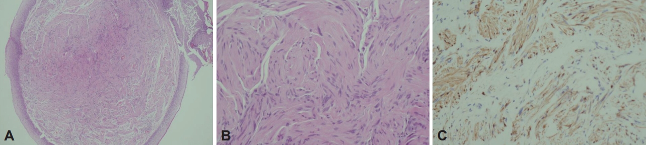
ĻĄ┤Ļ│ĪĒśĢ ļé┤ņŗ£Ļ▓Į ņ£ĀļÅäĒĢś ņāØĻ▓ĆņØĆ ĒÖśņ×ÉņØś ĻĄ¼ņŚŁ ļ░śņé¼Ļ░Ć ņŗ¼ĒĢśĻ│Ā ņóģļ¼╝ņØś Ēü¼ĻĖ░Ļ░Ć ņ×æņĢäņä£ ņŗżĒī©ĒĢśņśĆļŗż. ņĢĀņä▒ņØĆ ņä▒ļ¼ĖļČĆņĀäņŚÉ ņØśĒĢ£ Ļ▓āņ£╝ļĪ£ ņāØĻ░üļÉśņ¢┤ ņØīņä▒ņ╣śļŻī Ēøä Ļ▓ĮĻ│╝Ļ┤Ćņ░░ĒĢśĻĖ░ļĪ£ ĒĢśņśĆļŗż. Ēö╝ņŚ┤ Ļ░äĻĘ╣ņØś ņ£ĀļæÉņóģ ņØśņŗ¼ ņóģļ¼╝ņØĆ ņĪ░ņ¦üĒĢÖņĀü ĒÖĢņ¦ä ļ░Å ņØ┤ļ¼╝Ļ░É ĒśĖņĀäņØä ņ£äĒĢśņŚ¼ ņłśņłĀņØä Ļ│äĒÜŹĒĢśņśĆļŗż. ņ¦ĆņåŹņŖłĒŹ╝ĒÄäņŖż ļ░®ļ▓Ģņ£╝ļĪ£ 2.5ņÖĆĒŖĖ Ļ░ĢļÅäņØś CO2 ļĀłņØ┤ņĀĆ(AcuPulse; Lumenis Ltd. Yokneam, Israel) ļź╝ ņé¼ņÜ®ĒĢśņśĆļŗż. ĒśäņłśĒøäļæÉĻ▓ĮņØä Ļ▒░ņ╣śĒĢśņŚ¼ ļ│æļ│ĆņØä ļģĖņČ£ņŗ£ĒéżĻ│Ā(Fig. 2A), ņóģļ¼╝ņØś Ēīīņóģ ļ░®ņ¦Ćļź╝ ņ£äĒĢśņŚ¼ Ļ▓Ėņ×ÉļĪ£ ņ×Īņ¦Ć ņĢŖĻ│Ā ņóģļ¼╝ ņŻ╝ļ│ĆņŚÉ ļĀłņØ┤ņĀĆļź╝ ņĪ░ņé¼ĒĢśņŚ¼ ņĀÉļ¦ēĻ│╝ ņóģļ¼╝ņØä ļ░Ģļ”¼ĒĢśņśĆļŗż. ĻĘĖļ”¼Ļ│Āļéśņä£ ņóģļ¼╝ņØś Ļ░ĆļÅÖņä▒ņØä ĒÖĢņØĖĒĢ£ ņØ┤ĒøäņŚÉ Ļ▓Ėņ×ÉļĪ£ ņóģļ¼╝ņØĆ ņ×ĪĻ│Āņä£ ļĀłņØ┤ņĀĆļĪ£ ņĀłņĀ£ĒĢśņśĆļŗż. ņóģļ¼╝ ņŻ╝ļ│ĆņŚÉ ļ»ĖņäĖĒśłĻ┤ĆņØ┤ ļ░£ļŗ¼ĒĢśņŚ¼ ņČ£ĒśłņØ┤ ņ׳ņŚłņ£╝ļéś ņŚÉĒö╝ļäżĒöäļ”░ Ļ▒░ņ”łņÖĆ ļĀłņØ┤ņĀĆļź╝ ņØ┤ņÜ®ĒĢśņŚ¼ ņ¦ĆĒśłĒĢśĻ│Ā ņłśņłĀņØä ņóģļŻīĒĢśņśĆļŗż(Fig. 2B). ņłĀ Ēøä 1ņØ╝ņ¦ĖņŚÉ ļé┤ņŗ£Ļ▓Į ņåīĻ▓¼ņŚÉņä£ ņłśņłĀ ļČĆņ£äņØś ņČ£Ēśł, ļČĆņóģ, ĒØĪņØĖ ļ░Å ņä▒ļīĆ ņøĆņ¦üņ×ä ņĀĆĒĢś ļō▒ņØś ņåīĻ▓¼ņØĆ ņŚåņŚłĻ│Ā, ĒÖśņ×ÉļŖö ņØīņä▒ ņĢłņĀĢ ļ░Å ņŻ╝ņØśņé¼ĒĢŁ ĻĄÉņ£Ī Ēøä ļŗżņØīļéĀ Ēć┤ņøÉĒĢśņśĆļŗż(Fig. 1C). ņĪ░ņ¦üļ│æļ”¼ ņåīĻ▓¼ņØĆ hematoxylin and eosin (H&E) ņŚ╝ņāēņŚÉņä£ ĒÄĖĒÅēņäĖĒżĒĢś ņĪ░ņ¦üņŚÉ ņāØĻĖ┤ ņóģņ¢æņŚÉņä£ ņäĖĒżļ░ĆļÅäĻ░Ć ļŗżņ¢æĒĢśĻ│Ā ņĢĮĻ░äņØś ņØ┤ĒśĢņä▒ņØä ļ│┤ņØ┤ļŖö ļ░®ņČöĒśĢ ņäĖĒżĻ░Ć ļ¼╝Ļ▓░ ĒśĢĒā£ļĪ£ ņ”ØņŗØĒĢśļŖö ņåīĻ▓¼ņØ┤ņŚłĻ│Ā(Fig. 3A and B), ļ®┤ņŚŁņĪ░ņ¦üĒĢÖņŚ╝ņāē 200ļ░░ņŚÉņä£ S-100 ļŗ©ļ░▒ņ¦ł ņ¢æņä▒ ņåīĻ▓¼ņØ┤ Ļ┤Ćņ░░ļÉśņ¢┤ ņŗĀĻ▓Įņ┤łņóģņ£╝ļĪ£ ĒÖĢņ¦äļÉśņŚłļŗż(Fig. 3C).
ņØĖĒøäļæÉ ņØ┤ļ¼╝Ļ░ÉņØĆ ņłĀ Ēøä 2-3ņŻ╝ Ļ╣īņ¦ĆļŖö ņłĀ ņĀä ļ│┤ļŗż ļŹö ņĢģĒÖöļÉśņŚłļŗżĻ│Ā ĒĢśņśĆņ£╝ļéś, 4ņŻ╝ ļČĆĒä░ļŖö ĒśĖņĀäļÉśļŖö ņ¢æņāüņØ┤ņŚłļŗż. ļśÉĒĢ£ 1Ļ░£ņøö Ēøä ņŗ£Ē¢ēĒĢ£ ļé┤ņŗ£Ļ▓Į Ļ▓Ćņé¼ņŚÉņä£ļŖö ņłśņłĀ ļČĆņ£ä ņĀÉļ¦ēņØ┤ ņל ņ╣śņ£ĀļÉśĻ│Ā ĒŖ╣ņØ┤ ņåīĻ▓¼ņØĆ ņŚåņŚłļŗż(Fig. 1D). ņłĀ Ēøä 8ņŻ╝ņŚÉ ņŗ£Ē¢ēĒĢ£ ņØīņä▒Ļ▓Ćņé¼ņŚÉņä£ļŖö ņŻ╝ņÜö ĒÅēĻ░Ć ņ¦ĆĒæ£ņŚÉņä£ ņłĀ ņĀäĻ│╝ ņ£Āņé¼ĒĢ£ Ļ▓░Ļ│╝ļź╝ ļ│┤ņśĆņ£╝ļ®░ 8Ļ░£ņøöņØ┤ Ļ▓ĮĻ│╝ĒĢ£ Ēśäņ×¼Ļ╣īņ¦Ć ņ×¼ļ░£ņåīĻ▓¼ ņŚåņØ┤ ņČöņĀüĻ┤Ćņ░░ ņżæņØ┤ļŗż.
Ļ│Ā ņ░░
ņŗĀĻ▓Įņ┤łņóģņØĆ ņŗĀĻ▓Įņä¼ņ£ĀņØś ņŗĀĻ▓Įņ┤łĻ░Ć ņ׳ļŖö ļćīņŗĀĻ▓Į, ĻĄÉĻ░ÉņŗĀĻ▓Į ļ░Å ļ¦Éņ┤łņŗĀĻ▓ĮņØ┤ ņ׳ļŖö Ļ││ņŚÉņä£ ļ░£ņāØĒĢĀ ņłś ņ׳Ļ│Ā ņŗĀĻ▓Įņ┤łņóģņØś 25%-45%ņŚÉņä£ ļéśĒāĆļé£ļŗż[2]. ļæÉĻ▓ĮļČĆņŚÉņä£ļŖö ņ▓ŁņŗĀĻ▓ĮņØ┤ ĒśĖļ░£ ļČĆņ£äņØ┤ļ®░ ĻĘĖ ņÖĖņŚÉ ņØĖļæÉ ņŻ╝ņ£ä Ļ│ĄĻ░ä, ĒśĆ, ņØĖļæÉ, ņ×ģņłĀ ļ░Å ņŚ░ĻĄ¼Ļ░£ ļō▒ņŚÉņä£ ņל ļ░£ņāØĒĢ£ļŗż[2,6]. ĒøäļæÉ ņŗĀĻ▓Įņ┤łņóģņØĆ ņĀäņ▓┤ ņŗĀĻ▓Įņ┤łņóģņØś 0.1%-1.5%ļĪ£ ļō£ļ¼╝Ļ│Ā[7], ļīĆļČĆļČä Ēö╝ņŚ┤ĒøäļæÉĻ░£ņŻ╝ļ”äĻ│╝ Ļ░Ćņä▒ļīĆ ļō▒ņŚÉņä£ ļ░£ņāØĒĢśļ®░ 20ļīĆņŚÉņä£ 50ļīĆņŚÉ ņל ļ░£ņāØĒĢ£ļŗż[7]. ĒøäļæÉ ņŗĀĻ▓Įņ┤łņóģņØś ĻĖ░ņøÉņØĆ ņāüĒøäļæÉņŗĀĻ▓ĮņØś ļé┤ļČäņ¦ĆĻ░Ć Ļ░Ćņן ĒØöĒĢśņ¦Ćļ¦ī ļ░śĒÜīĒøäļæÉņŗĀĻ▓ĮņŚÉņä£ļÅä ĻĖ░ņøÉĒĢĀ ņłś ņ׳ņ£╝ļ®░ ļæÉ ņŗĀĻ▓ĮņŚÉņä£ ļÅÖņŗ£ņŚÉ ĻĖ░ņøÉĒĢśļŖö Ļ▓ĮņÜ░ļÅä ņ׳ļŗż[2,4,7].
ņóģņ¢æ ļ░£ņāØņØś Ļ░ĆņäżļĪ£ļŖö ņāüĒö╝ņäĖĒż ņ”ØņŗØņŚÉ ņØśĒĢ£ ļ╣äļ¦īņäĖĒżņØś ņ”ØĻ░Ć ļ░Å ņŗĀĻ▓ĮņåÉņāüņØ┤ļéś ņ×ÉĻĘ╣ņŚÉ ņØśĒĢ£ ņØ┤ņ░©ņĀü ļ░£ņāØ ļō▒ņØ┤ ņ׳ļŗż[6]. ņØĖĒøäļæÉ ņŗĀĻ▓Įņ┤łņóģņØś ņ×äņāü ņ¢æņāüņØĆ ļŗżņ¢æĒĢśņ¦Ćļ¦ī ņóģļ¼╝ņØ┤ ņ▓£ņ▓£Ē׳ ņä▒ņןĒĢśļ»ĆļĪ£ ļ¼┤ņ”ØņāüņØ┤ Ļ░Ćņן ĒØöĒĢśļ®░, ņóģļ¼╝ņØ┤ ņä▒ņןĒĢśļ®┤ ņĢĀņä▒, ĻĖ░ņ╣©, ņØĖĒøä ņØ┤ļ¼╝Ļ░É, ĒśĖĒØĪĻ│żļ×Ć ļ░Å ņŚ░ĒĢśĻ│żļ×Ć ļō▒ņØś ņ”ØņāüņØ┤ ļéśĒāĆļéĀ ņłś ņ׳ļŗż[2]. ņ¦äļŗ©ņØĆ ņ×äņāüņ¢æņāü, ņØ┤ĒĢÖņĀü Ļ▓Ćņé¼, ļé┤ņŗ£Ļ▓Į ļ░Å ņśüņāüĻ▓Ćņé¼ ņåīĻ▓¼Ļ│╝ ņäĖņ╣©ĒØĪņØĖĻ▓Ćņé¼ ļō▒ņ£╝ļĪ£ Ļ░Éļ│äņ¦äļŗ©ĒĢśĻ│Ā, ļ│æļ”¼ņĪ░ņ¦üĻ▓Ćņé¼ļź╝ ĒåĄĒĢśņŚ¼ ĒÖĢņ¦äļÉ£ļŗż[1,2]. ņØ╝ļ░śņĀüņØĖ ļé┤ņŗ£Ļ▓Į ņåīĻ▓¼ņØĆ ļ¦żļüłĒĢ£ Ēö╝ļ¦ēņ£╝ļĪ£ ļæśļ¤¼ņīōņØ┤ļ®░ ņĢĮĻ░äņØś Ļ▓░ņĀłņä▒ņØä Ļ░Ćņ¦ä ĻĄ¼ĒśĢ ļśÉļŖö ļé£ņøÉĒśĢ ļŗ©ņØ╝ņä▒ ņóģļ¼╝ļĪ£ ļ│┤ņØ┤Ļ│Ā, ņóģļ¼╝ņØ┤ ņä▒ņןĒĢśļ®┤ ļéŁĒżņä▒ ļ░Å ņäØĒÜīĒÖö ļ│Ćņä▒ ļō▒ņØś ļŗżņ¢æĒĢ£ ĒśĢĒā£ļĪ£ ļéśĒāĆļéĀ ņłś ņ׳ļŗż[6,8,9]. ļ│Ė ņ”ØļĪĆļŖö ņóģļ¼╝ņØś Ēü¼ĻĖ░Ļ░Ć ņ×æĻ│Ā Ēæ£ļ®┤ņØ┤ ņ£ĀļæÉņ¢æ ļ¬©ņŖĄņØ┤ Ļ┤Ćņ░░ļÉśņ¢┤ ņ£ĀļæÉņóģņ£╝ļĪ£ ņČöņĀĢĒĢśņśĆĻ│Ā, ĻĄŁņåīļ¦łņĘ©ĒĢś ņāØĻ▓ĆņØ┤ ņ¢┤ļĀżņøī ĒøäļæÉļ»ĖņäĖņłśņłĀļĪ£ ĒÖĢņ¦äļÉśņŚłļŗż.
ņŗĀĻ▓Įņ┤łņóģņØś ņĪ░ņ¦üļ│æļ”¼ņåīĻ▓¼ņØĆ H&E ņŚ╝ņāēņŚÉņä£ ļåÆņØĆ ļ░ĆļÅäļĪ£ ĻĄ¼ņä▒ļÉ£ ņäĖĒż ņśüņŚŁĻ│╝ ņäĖĒżĻ░Ć ļČłĻĘ£ņ╣ÖĒĢśĻ▓ī ļ░░ņŚ┤ļÉ£ ļé«ņØĆ ļ░ĆļÅäņØś ļČĆņóģ ņśüņŚŁņØ┤ Ēś╝ņ×¼ļÉ£ ņ¢æņāüņØä ļ│┤ņØ┤Ļ│Ā, ļ®┤ņŚŁņĪ░ņ¦üĒÖöĒĢÖ ņŚ╝ņāēņŚÉņä£ S-100 ļŗ©ļ░▒ņ¦łņŚÉ ņ¢æņä▒ņØä ļ│┤ņØĖļŗż[4,8]. ĒøäļæÉ ņŗĀĻ▓Įņ┤łņóģņØś ņ╣śļŻīļŖö ĒøäļæÉ ĻĖ░ļŖźņØä ļ│┤ņĪ┤ĒĢśļ®┤ņä£ ņóģņ¢æņØä ņÖäņĀäĒĢśĻ▓ī ņĀłņĀ£ĒĢśļŖö Ļ▓āņØ┤ļŗż[8]. Ēü¼ĻĖ░Ļ░Ć ņ×æņØĆ Ļ▓ĮņÜ░ļŖö CO2 ļĀłņØ┤ņĀĆļź╝ ņØ┤ņÜ®ĒĢ£ ĒøäļæÉļ»ĖņäĖņłśņłĀļĪ£ ņĀ£Ļ▒░ Ļ░ĆļŖźĒĢśņ¦Ćļ¦ī, Ēü░ Ļ▓ĮņÜ░ņŚÉļŖö ņĖĪņØĖļæÉ ņĀłĻ░£, ņÖĖņĖĪ Ļ░æņāüņŚ░Ļ│© ņĀłĻ░£ ļ░Å ņĀĢņżæ Ļ░æņāüņŚ░Ļ│© ņĀłĻ░£ ļō▒ņØä ĒåĄĒĢ£ ņÖĖļČĆ ņĀæĻĘ╝ļ▓ĢņØ┤ ĒĢäņÜöĒĢĀ ņłś ņ׳ļŗż[8,10].
ņĀĆņ×ÉļōżņØś Ļ▓Ćņāēņ£╝ļĪ£ļŖö ļ│Ė ņ”ØļĪĆņÖĆ Ļ░ÖņØ┤ Ēü¼ĻĖ░Ļ░Ć ņ×æĻ│Ā ņóģļ¼╝ Ēæ£ļ®┤ņØ┤ ņ£ĀļæÉņ¢æ ņ¢æņāüņØä ļ│┤ņØ┤ļŖö ņŗĀĻ▓Įņ┤łņóģņØĆ ļ│┤Ļ│ĀļÉ£ ļ░öĻ░Ć ņŚåļŗż. ļ│Ė ņ”ØļĪĆļź╝ ĒåĄĒĢ┤ņä£ ļ¦żņÜ░ ļō£ļ¼╝ņ¦Ćļ¦ī Ēö╝ņŚ┤Ļ░äĻĘ╣ņŚÉ ļ░£ņāØĒĢśļŖö ņ£ĀļæÉņ¢æ ņóģļ¼╝ņØś Ļ░Éļ│äņ¦äļŗ©ņŚÉ ņ£ĀļæÉņóģ ņÖĖņŚÉ ņŗĀĻ▓Įņ┤łņóģņØś Ļ░ĆļŖźņä▒ļÅä ņŚ╝ļæÉņŚÉ ļæÉņ¢┤ņĢ╝ ļÉ£ļŗżļŖö ĻĄÉĒøłņØä ņ¢╗ņØä ņłś ņ׳ņŚłļŗż.